Anatomy Of Musckes Sndctendons - Muscle Anatomy - Skeletal Muscles - Groin Muscles - Calf ... / A tendon connects the muscle to the bone.. New users enjoy 60% off. Lesson on the anatomy of the forearm: Muscles and tendons of upper leg. Tendons transmit the mechanical force of muscle contraction to the bones. Muscle anatomy chart 12 photos of the muscle anatomy chart abdominal muscle anatomy chart, human muscle anatomy diagram free, interactive muscle anatomy chart, pelvic muscle anatomy chart, shoulder muscle anatomy chart, human muscles, abdominal muscle anatomy chart, human muscle anatomy diagram free, interactive muscle.
Most of the muscles which act on the wrist joint are situated within the forearm, with only the tendon crossing the joint and inserting on the hand. The shoulder is not a single joint, but a complex arrangement of bones, ligaments, muscles, and tendons that is better called the shoulder girdle. *the origin, insertion, and belly.* a muscle's origin is where a tendon attaches it to the *less* movable bone. See tendons muscles foot lower leg anatomy stock video clips. Beranda anatomy of musckes sndctendons / upper limb muscle anatomy | 3d anatomy with actions of.
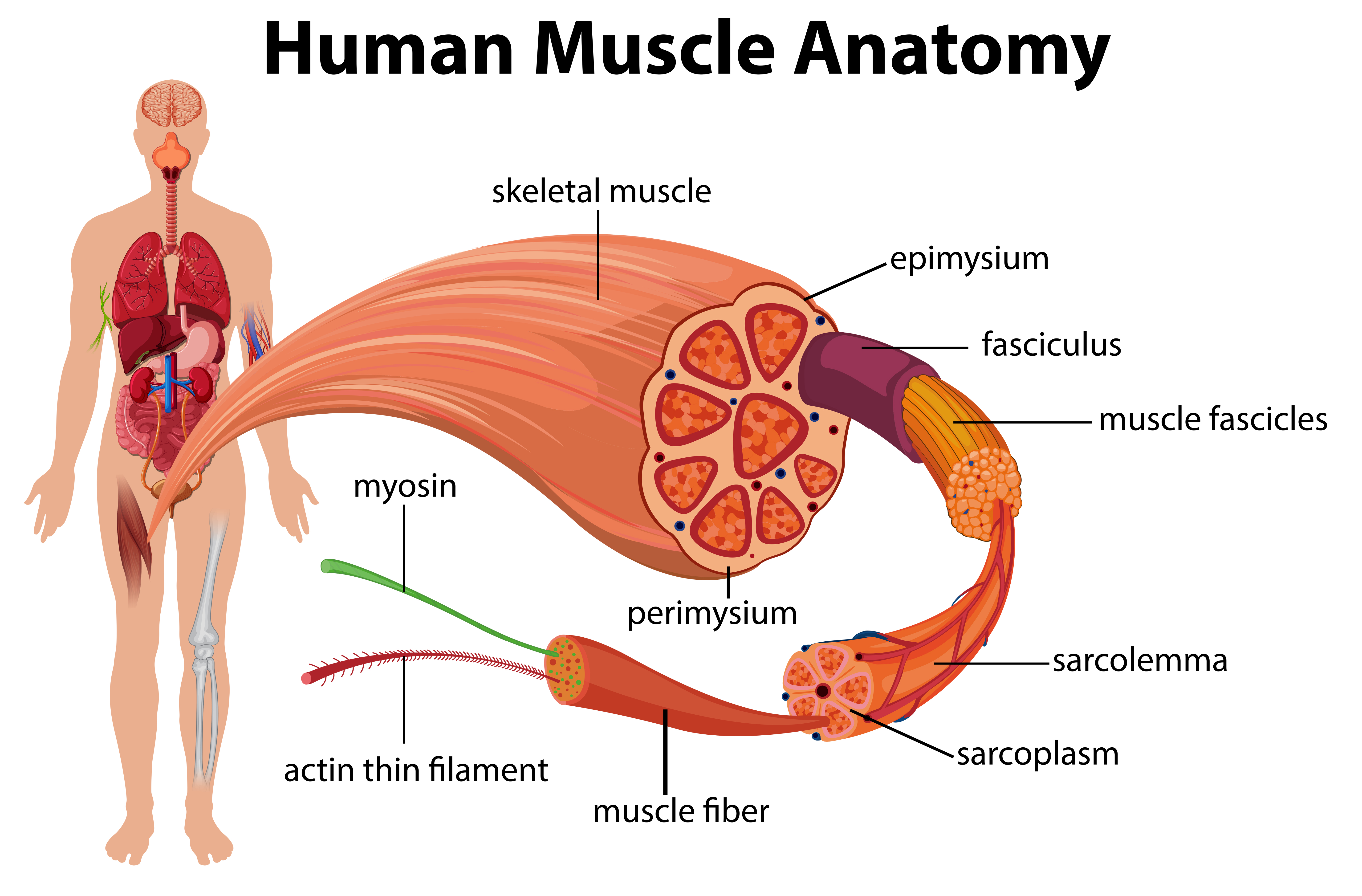
Menschliches Muskel-Anatomie-Diagramm - Download Kostenlos ... from static.vecteezy.com For example, the sternocleidomastoid muscle (neck area) assists with movement of the head, while the psoas major muscle (low back area) is associated with flexion of the thigh. Related posts of back muscles and tendons anatomy muscle anatomy chart. Tendons and ligaments are bands of connective tissue that help stabilize the body and allow movement. Most of the muscles which act on the wrist joint are situated within the forearm, with only the tendon crossing the joint and inserting on the hand. When the muscle contracts, the tendons are pulled, and the bone is moved. Tendons consist of densely packed collagen fibers. Muscles and tendons of upper leg. A tendon connects the muscle to the bone.
Major muscles of the ankle.
Major muscles of the ankle. Tendons and ligaments are bands of connective tissue that help stabilize the body and allow movement. *the origin, insertion, and belly.* a muscle's origin is where a tendon attaches it to the *less* movable bone. In this lesson, we look at the muscle. Learn about the anatomy and physiology of tendons. Tendons consist of densely packed collagen fibers. Muscles, either individually or in groups, are supported by fascia. These muscles are similar to the thenar muscles in both name and organisation. In humans, the foot is one of the most complex structures in the body. As these muscles contract and relax, they move skeletal bones to create movement of the body. This is lesson 1 on the anatomy of the forearm. Every skeletal muscle has three main parts: Muscle anatomy chart 12 photos of the muscle anatomy chart abdominal muscle anatomy chart, human muscle anatomy diagram free, interactive muscle anatomy chart, pelvic muscle anatomy chart, shoulder muscle anatomy chart, human muscles, abdominal muscle anatomy chart, human muscle anatomy diagram free, interactive muscle.
*the origin, insertion, and belly.* a muscle's origin is where a tendon attaches it to the *less* movable bone. Ebraheim's educational animated video describes the muscle anatomy of the hip and buttocks region with simple images; These muscles allow the ankle to bend downward and outward. Every skeletal muscle has three main parts: Beranda anatomy of musckes sndctendons / upper limb muscle anatomy | 3d anatomy with actions of.
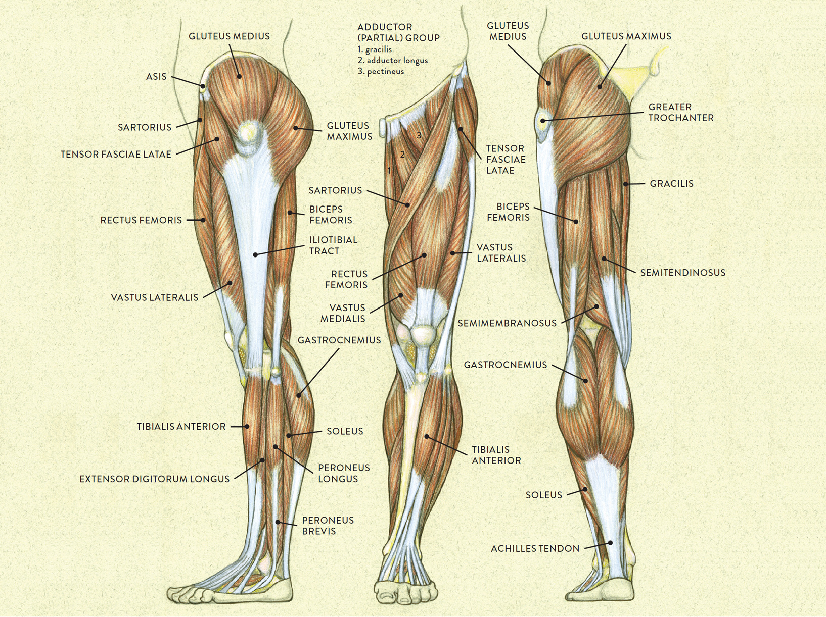
Gluteus anatomy and 4 effective exercises for your training from www.technogym.com The smaller bone that runs alongside the tibia (fibula) and the kneecap (patella) are the other bones that make the knee joint. This video also provides you with a. Tendons connect the knee bones to the leg muscles that move the knee. The shoulder is not a single joint, but a complex arrangement of bones, ligaments, muscles, and tendons that is better called the shoulder girdle. Download 4,505 human body muscles anatomy stock illustrations, vectors & clipart for free or amazingly low rates! Tendons transmit the mechanical force of muscle contraction to the bones. Tendons and ligaments are bands of connective tissue that help stabilize the body and allow movement. Learn about the muscles, tendons, bones, and ligaments that comprise the knee joint anatomy.
When muscles contract, they pull on the tendons to move the bones.
All together they help hold your upper arm in place in the shoulder. The hands enable us to perform many of our daily activities such as driving, writing and cooking. When muscles contract, they pull on the tendons to move the bones. The upper arm is located between the shoulder joint and elbow joint. For example, the sternocleidomastoid muscle (neck area) assists with movement of the head, while the psoas major muscle (low back area) is associated with flexion of the thigh. The knee joint is most significantly affected by two major muscle groups: Anatomy ankle anatomy ankle + ligament + tendon the foot anatomy human ankle anatomy 3d leg muscle lower leg anatomy leg articulation peroneal ankle muscles foot. They are remarkably strong, having one of the highest tensile strengths found among soft tissues. Every skeletal muscle has three main parts: The foot is a part of vertebrate anatomy which serves the purpose of supporting the animal's weight and allowing for locomotion on land. Muscles, either individually or in groups, are supported by fascia. Most of the muscles which act on the wrist joint are situated within the forearm, with only the tendon crossing the joint and inserting on the hand. Similar to ligaments, they are made of collagen and can withstand increased tension.
Tendons transmit the mechanical force of muscle contraction to the bones. The hands enable us to perform many of our daily activities such as driving, writing and cooking. Tendons connect the knee bones to the leg muscles that move the knee. Related posts of muscles and tendons of the leg muscle anatomy and physiology. Skeletal muscles are attached to bones by tendons and can be as long as 30 cm, although they are usually 2 to 3 cm in length.
LEFT: Lateral view from schoolbag.info Download 4,505 human body muscles anatomy stock illustrations, vectors & clipart for free or amazingly low rates! This is lesson 1 on the anatomy of the forearm. New users enjoy 60% off. Extensor carpi radialis brevis extensor carpi radialis longus Muscles, either individually or in groups, are supported by fascia. The peroneal muscles (peroneus longus and peroneus brevis), on the outside edge of the ankle and foot. Learn about the anatomy and physiology of tendons. Lying exposed between the protective bones of the superiorly located ribs and the inferiorly located pelvic girdle, the muscles of this region play a critical role in protecting the.
It also helps you raise and rotate your arm.
When the muscle contracts, the tendons are pulled, and the bone is moved. The fleshy, thick part of the muscle is called its belly. The knee joint is most significantly affected by two major muscle groups: Every skeletal muscle has three main parts: See tendons muscles foot lower leg anatomy stock video clips. For example, the sternocleidomastoid muscle (neck area) assists with movement of the head, while the psoas major muscle (low back area) is associated with flexion of the thigh. In this lesson, we look at the muscle. The human hand is made up of the wrist, palm, and fingers and consists of 27 bones, 27 joints, 34 muscles, over 100 ligaments and tendons, and many blood vessels and nerves. Extensor carpi radialis brevis extensor carpi radialis longus The muscles of the abdomen, lower back, and pelvis are separated from those of the chest by the muscular wall of the diaphragm, the critical breathing muscle. Anatomy ankle anatomy ankle + ligament + tendon the foot anatomy human ankle anatomy 3d leg muscle lower leg anatomy leg articulation peroneal ankle muscles foot. These muscles allow the ankle to bend downward and outward. Specific muscles are associated with specific movements of parts of the anatomy.